Superior Vena Cava
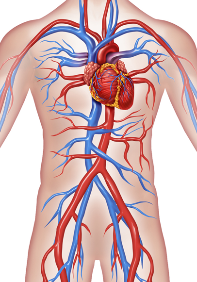
Superior vena cava is a vein of a very large diameter, yet is very short in length. It is the vein that drains deoxygenated blood in the right atrium (one of the four chambers) of the heart.
read moreSuperior Vena Cava
ANATOMICAL FEATURES
Superior vena cava is situated in the superior mediastinum (space in the chest between the two lungs). It is formed by the union of the right and left brachiocephalic veins, which receives blood from the eye, neck and the upper limbs. It starts at the level of the lower margin (border) of the first right costal (rib) cartilage. It is joined by the azygos vein just before it enters the right atrium. There is no valve present in the superior vena cava, as a result some blood is pushed into the internal jugular vein when the right atrium contracts.
FUNCTION
Superior vena cava carries deoxygenated blood from the upper half of the body and drains it into the heart’s right atrium.
CLINICAL IMPORTANCE
Superior vena cava can sometimes get blocked and result in a group of symptoms known as superior vena cava syndrome. This may be caused by lung cancer or NHL (Non-Hodgkin lymphoma). The symptoms can be difficulty in breathing, coughing or chest pain. It is treated by radiotherapy, surgery or stent placement.
Report Error


