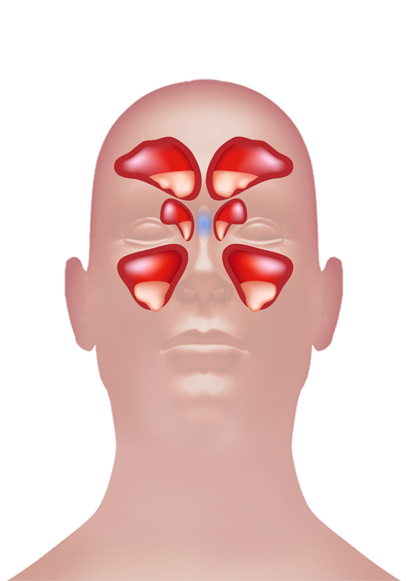
Maxillary Sinus
Sinus maxilliaris is the Latin pronunciation for the Maxillary Sinus. It is pyramidal in shape and is one of the largest Paranasal Sinuses. It drains into the Middle Meatus of Nose.
read more Sinus Frontalis Superior Nasal ConchalInferior Nasal Concha Nasal CavitySinus Sphenoidalis Alveoli Larynx Nasopharynx
Oropharynx Laryngopharynx Frontal SinusSphenoid SinusEthmoid Sinus Maxillary SinusBronchus Vertebrate Trachea Bronchioles Capillary Beds
Alveolar Duct Alveolar Sacs Pulmonary VeinPulmonary Artery
Maxillary Sinus
ANATOMY
Maxillary sinus is found in the Body of Maxilla and has three recesses which are as follows.
-
Alveolar Recess; it is pointed inferiorly and is bounded by the Maxillary Alveolar process.
-
Zygomatic Recess; bounded by the Zygomatic bone it is pointed laterally.
-
Infraorbital recess; it is bounded by the Interior Orbital Surface of the Maxilla and is pointed superiorly.
Medial Wall of the Maxillary sinus is formed of Cartilage. The Ostia (small opening) for the drainage are located high on the Medial Wall opening into Semilunar Hiatus of the lateral side of the Nasal Cavity. The position of Osita prevents the drainage of Maxillary sinus contents in the head when it is erect. The ostium of the Maxillary sinus is of 2.4 mm in diameter with a volume of 10ml located high up on the Medial Wall. Maxillary Sinus is lined with Mucoperiosteum also known as Schneiderian Membrane. This membrane is made up of Ciliated Columnar Epithelial Cells on the inside and Periosteum on the outside. The posterior wall of the Maxillary sinus transmits posterior superior alveolar nerves and vessels to the Molar Teeth. The floor of Maxillary Sinus is formed by the Alveolar process of the Maxilla.
BLOOD SUPPLY
The three arteries that supply Maxillary sinus are as follows.
-
Posterior superior Alveolar Artery
-
Infraorbital Artery
-
Posterior Nasal Orbital Artery
These three arteries are branches of Maxillary Artery.
INNERVATION
For mucous secretions the Mucous Membrane receives their innervations from the Postganglionic Parasympathetic Nerves which originates from the branches of Great Petrosal Nerve. Sensory innervations are provided from the branches of Maxillary Nerve i.e. Superior Alveolar Nerves.
DEVELOPMENT
It is developed throughout the childhood, before then at time of birth it is only as Rudimentary Air cells.
FUNCTION
Maxillary Sinus plays a key role by reducing the weight of the Cranium, performing functions of the Resonate Bone and controls the inhaled air temperatures.
CLINICAL SIGNIFICANCE
MAXILLARY SINUSITIS
The most common disease of the Maxillary Sinus is the Maxillary Sinusitis due to inflammation. The symptoms of it are Headache near Sinus, pharyngeal discharge, fever and weakness. The main for Sinusitis is dentally related.
CANCER
Carcinoma of the Sinuses may invade the Palate and cause dental pain. It can also block the Nasolacrimal duct.
Report Error


